ACTIVA WORKS
A Swedish study contradicts the tremendous success that thousands of practitioners have experienced with millions of ACTIVA restorations over the past six years. The investigator had no clinical experience with ACTIVA, did not follow written instructions for etching and bonding, and did not make cavity preps with the intention of creating retention form. A bonding agent was not used in any of the cases. Pulpdent always recommends a bonding agent with ACTIVA when there is insufficient retention form. Lost restorations reported in the study are proof of non-retentive cavity preps.
Modern approaches to restorative dentistry may threaten conventional methods and materials. However, moisture-friendly resins that adapt intimately to tooth structure and support the natural remineralization process have the potential to elevate our practice of dentistry, extend the life of composite restorations, and benefit the health and welfare of our patients.
The results of both clinical and in vitro studies over a period of 6-8 years, along with the experience of thousands of dentists worldwide, cannot be denied. ACTIVA is esthetic, strong, durable, tough, and wear and fracture resistant. Margins remain imperceptible and without staining over time. Clinicians note the excellent condition of the soft tissues. The release and recharge of calcium, phosphate and fluoride support biomineralization. ACTIVA WORKS!
Five Years of Success
ACTIVA BioACTIVE: Composite resin with high phosphate release, a true glass ionomer reaction, and rubberized-resin component. Highly esthetic, tough, durable, resists wear, fracture and chipping, moisture friendly, for all restorative indications. Mimics the slow, natural biomineralization process.
After 7 million (and counting) successful clinical placements, we know Activa Works
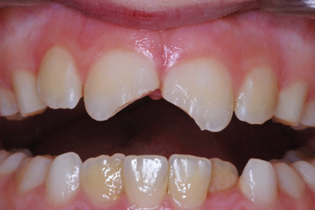
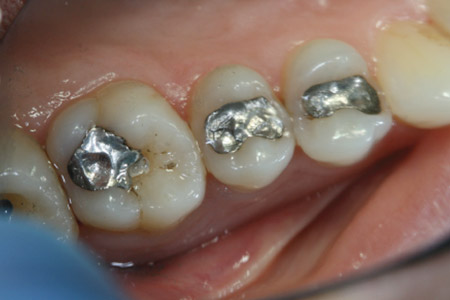
Before
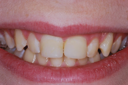
39-Month Recall
Before
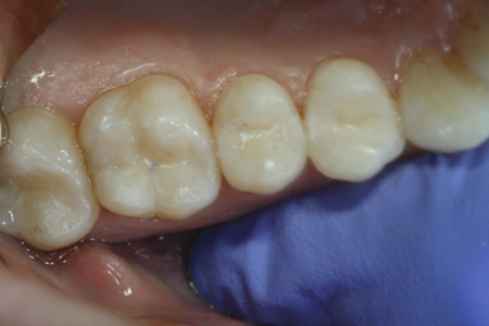
29-Month Recall
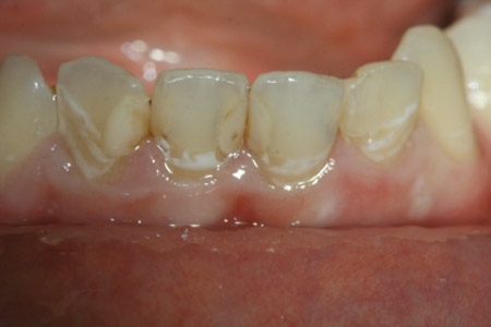
Before
37-Month Recall
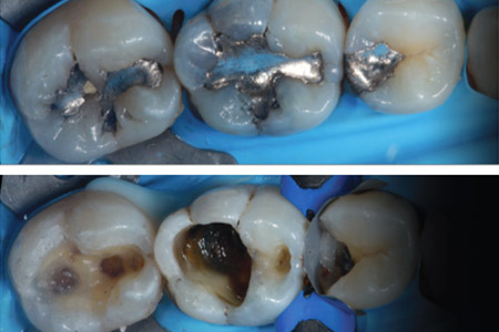
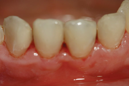
Before
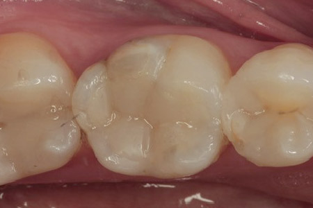
42-Month Recall
What Clinicians and Researchers are Saying about ACTIVA
“98% rating at recall [after two years]. Retention was excellent.”
“No gap was observed between ACTIVA BioACTIVE and the tooth. Good integration and mineral formation was apparent.”
“ACTIVA showed significantly lower demineralization values in dentin margins, compared to the composite resins TE and SE [total-etch and self-etch], regardless of the adhesive system used.”
Research shows that biofilm is more easily removed from ACTIVA than from conventional composites.
“After close to 4 years of use, ACTIVA has proved to be durable and reliable.”
“Clinical observations confirm research findings regarding ACTIVA’s wear resistance and resistance to fracture.”
“ACTIVA had significantly less enamel demineralization adjacent to the bracket when compared to Transbond XT.”
Ions released from ACTIVA pass through the bonding agent. There is a lower concentration of MMPs with ACTIVA. [MMPs are a leading cause of composite restoration failure.]
Clinical Validation and Awards
Independent studies indicate that ACTIVA is tougher and more fracture resistant than all other direct restorative materials tested. ACTIVA’s patented rubberized-resin component resists wear, fracture and chipping. University testing shows that ACTIVA’s compressive strength is comparable to the leading composites and far superior to glass ionomers and RMGIs.
Wear and Toughness

Materials Tested: Dyract, CompGlass, ACTIVA Restorative, Beautifil-II, Fuji II LC
“ACTIVA Restorative showed significantly lower wear depth than all other tested materials.”
“ACTIVA Restorative exhibited the highest fracture toughness.”
Garoushi S, Vallittu PK, Lassila L. Characterization of flouride releasing restorataive dental materials. Dent Mater J
2018;37(2):293-300.
A one-year clinical performance report from The Dental Advisor gives ACTIVA BioACTIVE-RESTORATIVE a 5-star rating. Ninety-six restorations recalled after one year were rated for lack of postoperative sensitivity, esthetics, resistance to fracture and chipping, resistance to marginal discoloration, wear resistance and retention. ACTIVA received a 98% clinical performance rating.

“I have used Activa BioActive-Restorative as a core in teeth with deep decay – the uptake of Ca, P and Fl could have a positive effect in these cases.”
“The esthetics were excellent at 2 years.”
ACTIVA BioACTIVE-RESTORATIVE performed extremely well at the two-year recall. Each evaluated category received a 96% or better rating. Overall, ACTIVA BioACTIVE-RESTORATIVE received a clinical performance rating of 98% at the two-year recall.
The BioACTIVE Difference
While traditional composites are passive in nature and have no bioactive potential, bioactive materials play a dynamic role in the mouth. They are moisture friendly and react to pH changes in the oral environment to help fortify and recharge the ionic properties of saliva, teeth and the material itself.
ACTIVA releases and recharges calcium, phosphate and fluoride. It stimulates apatite formation at the material-tooth interface. This natural remineralization/regenerative process knits the material and the tooth together, penetrates and fills micro-gaps, guards against secondary caries, and seals margins against microleakage and failure. This bioactive difference supports a prevention model and helps maintain the health of the dentition.
Understanding ACTIVA: Ensuring Success
Understanding Activa
- ACTIVA is moisture friendly. Only hydrophilic materials can be bioactive.
- ACTIVA releases and recharges calcium, phosphate and fluoride and supports the natural remineralization process.
- MMP reduction is reported with ACTIVA. MMPs degrade the adhesive layer and are the primary cause of restoration failure.
- Calcium, phosphate and fluoride ions diffuse through the bonding agents tested, supporting bioactivity and apatite formation.
- ACTIVA contains phosphate acid groups. It is rich in phosphates.
- ACTIVA contains a rubberized-resin molecule that resists fracture and chipping.
- ACTIVA combines esthetics and durability with bioactive properties.
ACTIVA + BONDING AGENT: MMPs and the ADHESIVE LAYER
- Ions released from ACTIVA pass through the bonding agent.
- ACTIVA prevents the breakdown of the bonding layer when the bonding agent is applied using the etch and rinse technique.
- This study indicates a lower concentration of MMPs with ACTIVA. MMPs are a leading cause of composite restoration failure.

Sauro S. Universidad CEU-Cardenal Herrera, Valencia, Spain 2018
ACTIVA + BONDING AGENT
Ions released from ACTIVA pass through the bonding agent

This study demonstrated that fluoride ions from a restorative material are able to penetrate through the adhesive bonding agents tested.
Murali S, Epstein N, Perry R, Kugel G.
Tufts University

- ACTIVA BioACTIVE-RESTORATIVE was able to form a crystalline mineral at the tooth-material interface after 30 days in simulated saliva solution.
- Adhese Universal (Ivoclar-Vivadent) was used as the bonding agent.
Lloyd JL, Freitas H, Comisi JC.
Medical University of South Carolina 2019
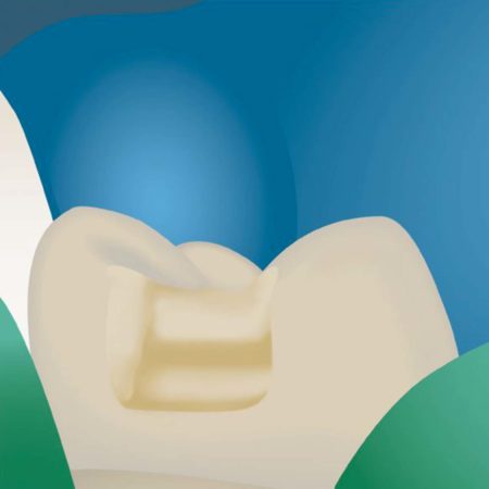
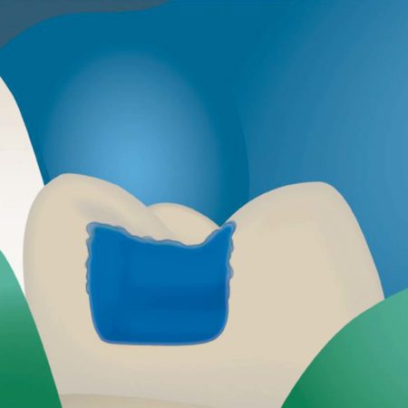
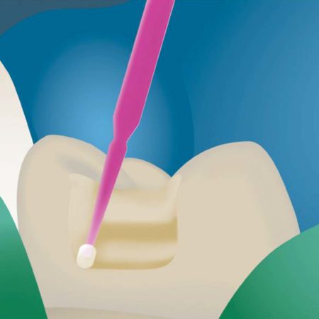
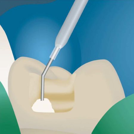
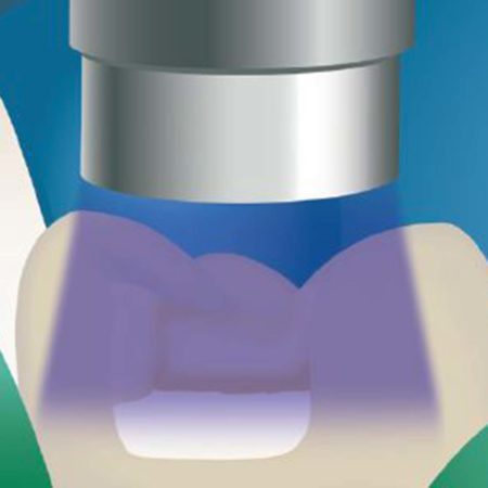
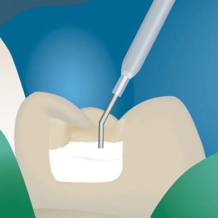
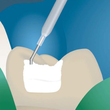
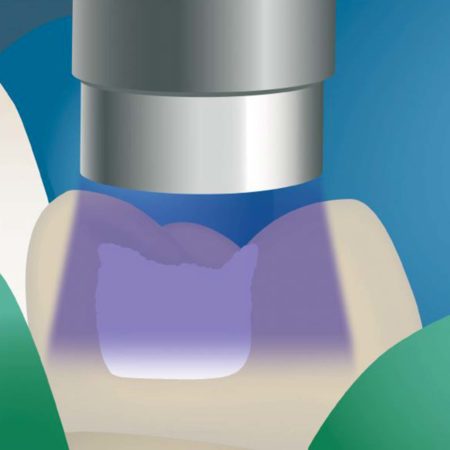
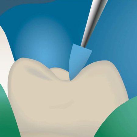
Instructions For Use
ACTIVA References
Fluoride ion release and recharge over time in three restoratives. Slowokowski L, et al. J Dent Res 93 (Spec Iss A) 268, 2014 (iadr.org).
Zmener O, Pameijer CH, Hernandez S. Resistance against bacterial leakage of four luting agents used for cementation of complete cast crowns. Am J Dent 2014;27(1):51-55.
Zmener O, Pameijer CH, et al. Marginal bacterial leakage in class I cavities filled with a new resin-modified glass ionomer restorative material. 2013.
Flexural strength and fatigue of new Activa RMGIs. Garcia-Godoy F, et al. J Dent Res 93 (Spec Iss A) 254, 2014 (iadr.org).
Deflection at break of restorative materials. Chao W, et al. J Dent Res 94 (Spec Iss A) 2375, 2015 (iadr.org).
McCabe JF, et al. Smart Materials in Dentistry (abst). Aust Dent J 2011 56 Suppl 1:3-10.
Pilot study to measure fluoride ion penetration of hydrophilic sealant. Cannon M, et al. J Dent Res 89 (Spec Iss A) 1345, 2010 (www.iadr.org).
Water absorption properties of four resin-modified glass ionomer base/liner materials. (Pulpdent)
pH dependence on the phosphate release of Activa ionic materials. (Pulpdent)
Kane B, et al. Sealant adaptation and penetration into occlusal fissures. Am J Dent 2009;22(2):89-91.
Ion release from a new protective coating. Rusin RP, et al. J Dent Res 88 (Spec Iss A) 3200, 2009 (www.iadr.org).
Comparison of antimicrobial properties of sealants and amalgam. Sharma S, Kugel G, et al. J Dent Res 87 (Spec Iss B) 0450, 2008 (www.iadr.org).
Naorungroj S, et al. Antibacterial surface properties of fluoride-containing resin-based sealants. J Dent 2010.
Prabhakar AR, et al. Comparative evaluation of the length of resin tags, viscosity and microleakage of pit and fissure sealants – an in vitro SEM study. Contemp Clin Dent 2011;2(4):324-30.
Pameijer CH. Microleakage of four experimental resin modified glass ionomer restorative materials. April 2011.
Microleakage of dental bulk fill, conventional and self-adhesive composites. Cannavo M, et al. J Dent Res 93 (Spec Iss A) 847, 2014 (iadr.org).
Comparison of Mechanical Properties of Dental Restorative Material. Girn V, et al. J Dent Res 93 (Spec Iss A) 1163, 2014 (iadr.org).
Mechanical properties of four photo-polymerizable resin-modified baseliner materials. (Pulpdent)
Singla R, et al. Comparative evaluation of traditional and self-priming hydrophilic resin. J Conserv Dent 2012;15(3):233-6.
Water absorption and solubility of restorative materials. (Pulpdent)
Increasing the Service Life of Dental Resin Composites. www.nidcr.nih.gov. grants & funding. concept clearances. May 2009.
Spencer P, et al. Adhesive dentin interface the weak link in the composite restoration. Am Biomed Eng 2010;38(6):1989-2003.
Murray PE, et al. Analysis of pulpal reactions to restorative procedures, materials, pulp capping, and future therapies. Crit Rev Oral Biol Med 2002;13:509.
DeRouen TA, et al. Neurobehavioral effects of dental amalgam in children a randomized clinical trial. JAMA 2006;295(15):1784-1792.
Nordbo H, et al. Saucer-shaped cavity preparations for posterior approximal resin composite restorations observations up to 10 years. Quintessence Int 1998;29(1):5-11.
Skartveit L, et al. In vivo fluoride uptake in enamel and dentin from fluoride-containing materials. J Dent Child 1990; 57(2):97-100.
Wear of a calcium, phosphate and fluoride releasing restorative material. Bansal R, et al. J Dent Res 94 (Spec Iss A) 3797, 2015 (iadr.org).
Wear resistance of new ACTIVA compared to other restorative materials. Garcia- Godoy F, Morrow BR. J Dent Res 94 (Spec Iss A) 3522, 2015 (iadr.org).
Pameijer CH, Garcia-Godoy F, Morrow BR, Jefferies SR. Flexural strength and flexural fatigue properties of resin-modified glass ionomers. J Clin Dent 2015;26(1):23-27
Pameijer CH, Zmerner O, Kokubu G, Grana D. Biocompatibility of four experimental formulations in subcutaneous connective tissue of rats. 2011.
Pameijer CH, Zmener O. Histopathological evaluation of an RMGI ionic-cement [Pulpdent Activa], auto and light cured – A subhuman primate study. March 2011.
ACTIVA BioActive-Restorative 6-month clinical performance report. The Dental Advisor 2015. dentaladvisor.com.
ACTIVA BioActive-Restorative one-year clinical performance +++++. The Dental Advisor 2015. dentaladvisor.com.
Compressive strength and deflection at break of four cements. Daddona J, Pagni S, Kugel G. J Dent Res 95 (Spec Iss A) 0658, 2016 (iadr.org).
Surface deposition analysis of bioactive restorative material and cement. Chao W, Perry R, Kugel G. J Dent Res 95 (Spec Iss A) S1313, 2016 (iadr.org).
Comparison of compressive strength of liner materials. Epstein N, et al. J Dent Res 95 (Spec Iss A) S0653, 2016 (iadr.org).
Water absorption and solubility of four dental cements. Hall J, et al. J Dent Res 95 (Spec Iss A) S1126, 2016 (iadr.org).
Shear bond strength of several dental cements. Tran A, et al. J Dent Res 95 (Spec Iss A) S0579, 2016 (iadr.org).
Repetitive deflection strengths of adhesive cements. Samaha S, et al. J Dent Res 95 (Spec Iss A) S1076, 2016 (iadr.org).
Fluoride Release of Bioactive Restorative with Bonding Agents. Murali S, et al. J Dent Res 95 (Spec Iss A) S0368, 2016 (iadr.org).
Profilometry bioactive dental materials analysis and evaluation of dentin integration. Garcia-Godoy F, Morrow BR. J Dent Res 95 (Spec Iss A) 1828, 2016 (iadr.org).
Staining and whitening products induce color changes of multiple composites. Parks H, Morrow BR, Garcia-Godoy F. J Dent Res 95 (Spec Iss A) S1323, 2016 (iadr.org).
Profilometry based composite abrasion using different current dentifrices. Lindsay AA, Morrow BR, Garcia-Godoy F. J Dent Res 95 (Spec Iss A) S0318, 2016 (iadr.org).
Bansal R, Burgess JO, Lawson NC. Wear of an enhanced resin-modified glass-ionomer restorative material. Am J Dent 2016;29(3):171-174.
Evaluation of pH, fluoride and calcium release for dental materials. Morrow BR, Brown J, Stewart CW, Garcia-Godoy F. J Dent Res 96 (Spec Iss A) 1359, 2017 (iadr.org).
Adhesion of s. mutans biofilms on potentially antimicrobial dental composites. Mah J, Merritt J, Ferracane J. J Dent Res 96 (Spec Iss A) 2560, 2017 (iadr.org).
Microleakage under class ll restorations restored with bulk-fill materials. Kulkami P, et al. J Dent Res 96 (Spec Iss A) 2604, 2017 (iadr.org).
Fluoride release of dental restoratives when brushed with fluoridated toothpaste. Epstein N, Roomian T, Perry R. J Dent Res 96 (Spec Iss A) 1254, 2017 (iadr.org).
ACTIVA Bioactive-Restorative. Two-year clinical performance +++++. The Dental Advisor 2017, dentaladvisor.com.
May E, Donly KJ. Fluoride release and re-release from a bioactive restorative material. Am J Dent 2017;30(6):305-308.
Garoushi S, Vallittu PK, Lassila L. Characterization of fluoride releasing restorative dental materials. Dent Mater J 2018;37(2):293-300.
Reznik J, Kulkarni P, Shah S, Chang B, Burgess JO, Robles A, Lawson NC. Crown Retention Strength and Ion Release of Bioactive Cements. J Dent Res 97 (Spec Iss A) 656, 2018 (iadr.org).
Boutsiouki C, Lücker S, Domann E, Krämer N. Is a bioactive composite able to inhibit secondary caries. Justus-Liebig-Universitat Giessen, Vaterstetten. Germany 2017.
Alrahlah A. Diametral tensile strength, flexural strength, and surface microhardness of bioactive bulk fill restorative. J Contemp Dent Practice 2018;19(1):13-19.
Influence of novel bioactive materials on dentinal enzymatic activity. Comba A, Breschi L, et al. J Dent Res 97 (Spec Iss A) 0273, 2018 (iadr.org).
Dentifrices, surface roughness and depth loss of restorative materials. Smith JB, Lambert AN, Morrow BR, Pameijer CH, Garcia-Godoy F. J Dent Res 97 (Spec Iss A) 1621, 2018 (iadr.org).
Enamel demineralization adjacent to orthodontic brackets bonded with Active Bioactive Restorative. Saunders KG, Donley KJ, Mattevi G. of Texas Health Science Center, San Antonio 2017.
Bioactive materials, demineralization, and shear strength of orthodontic brackets. Donohue J, et al. J Dent Res 96 (Spec Iss A) 3289, 2017 (iadr.org).
Roulet J-F, et al. In vitro wear of two bioactive composites and a glass ionomer cement. DZZ International 2019;1(1):24-30.
Banon R, et al. Clinical evaluation of a new bioactive ionic resin material (ACTIVA™ BIOACTIVE) in primary molars – a split mouth randomized trial. Ghent University 2018.
Omidi BR, et al. Microleakage of an enhanced resin-modified glass ionomer restorative material in primary molars. Researchgate 2018;15(4)205-213.
Croll TP, Lawson NC. Activa Bioactive-Restorative material in children and teens: examples and 46-month observations. Inside Dentistry 2018.
Sauro S, et al. Effects of ions-releasing restorative materials on the dentine bonding longevity of modern universal adhesives after load-cycle and artificial saliva aging. Materials 2019;12:722.
Lloyd VJ, Hunter F, Comisi J. The bio-mineralization potential of various bioactive restorative materials, MUSC 2019.


